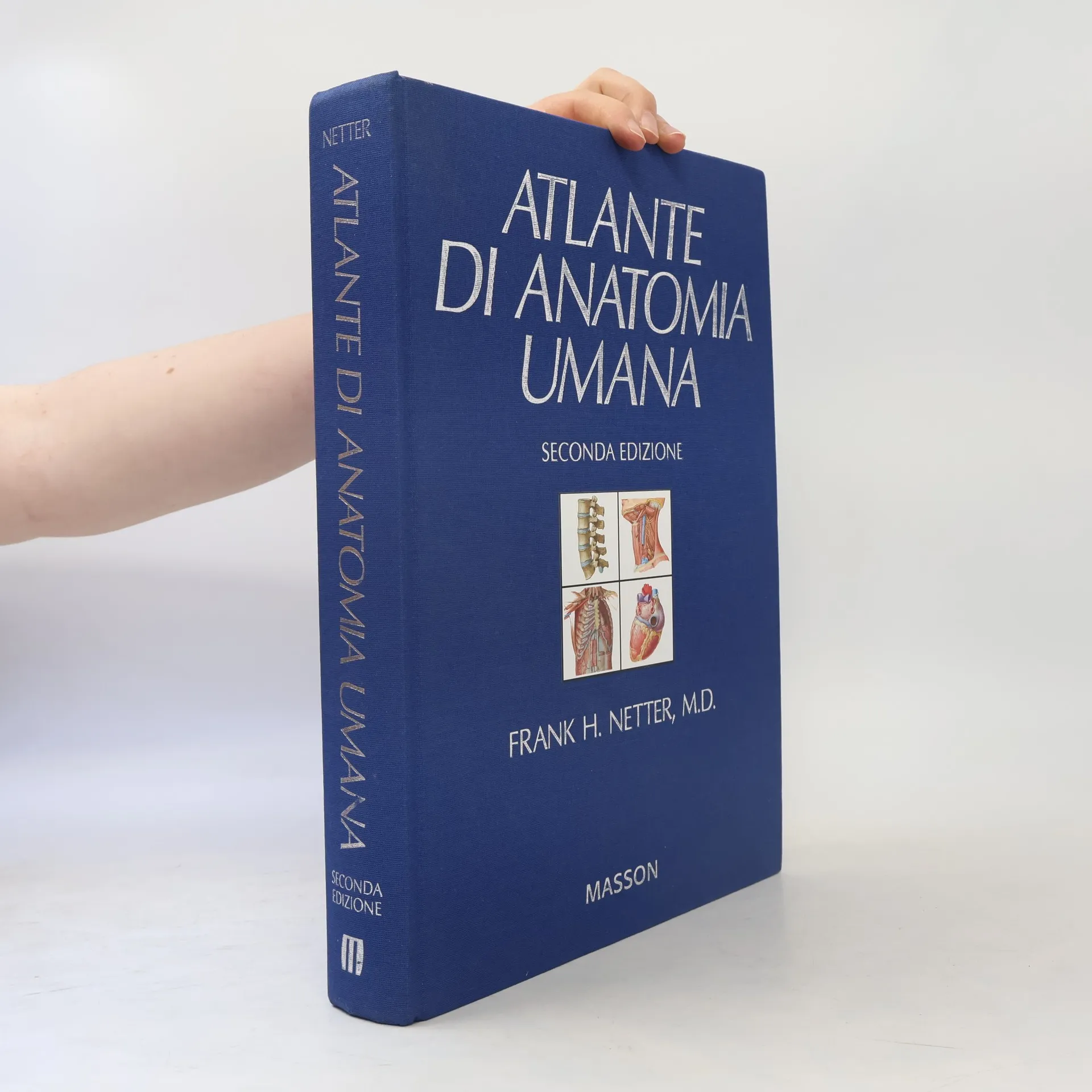
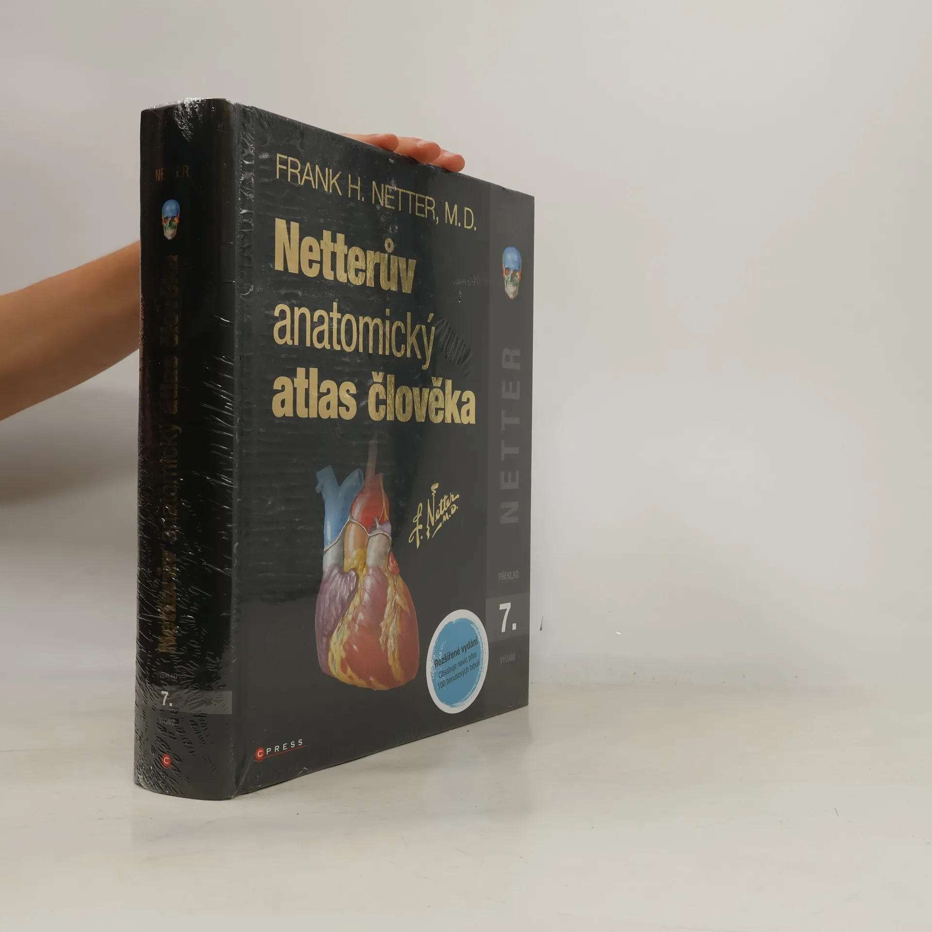
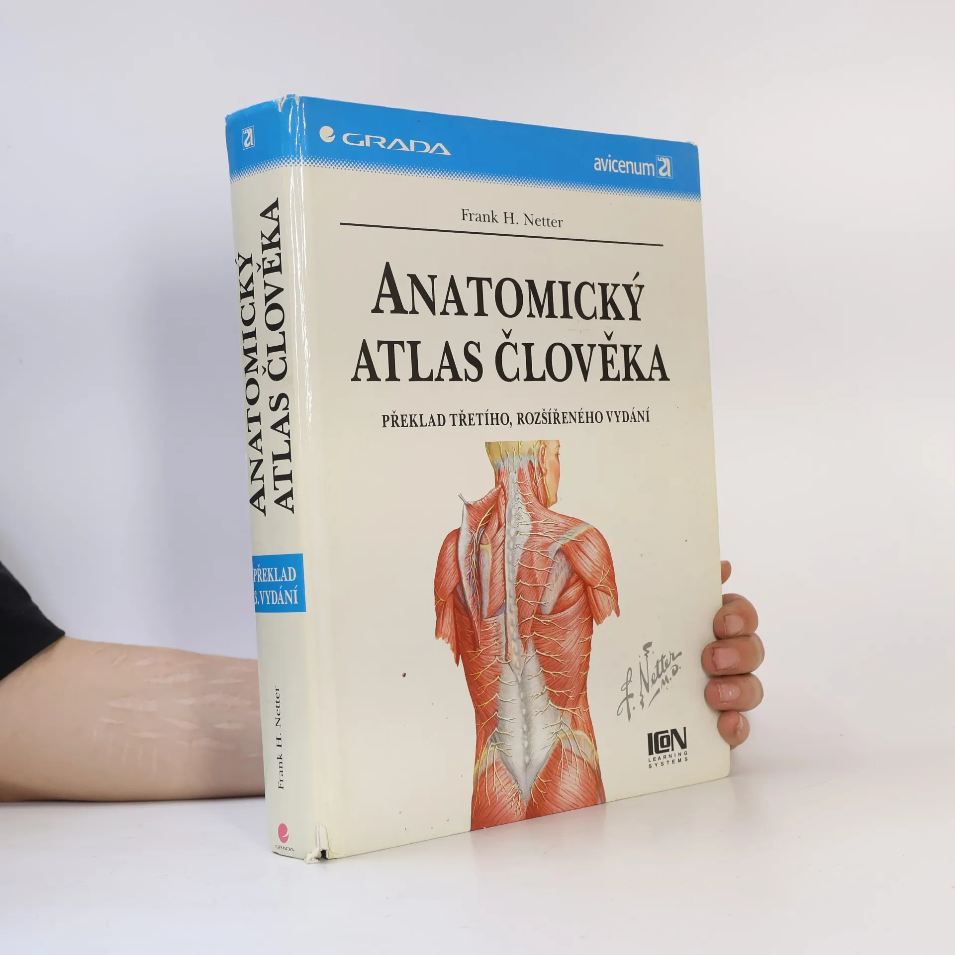
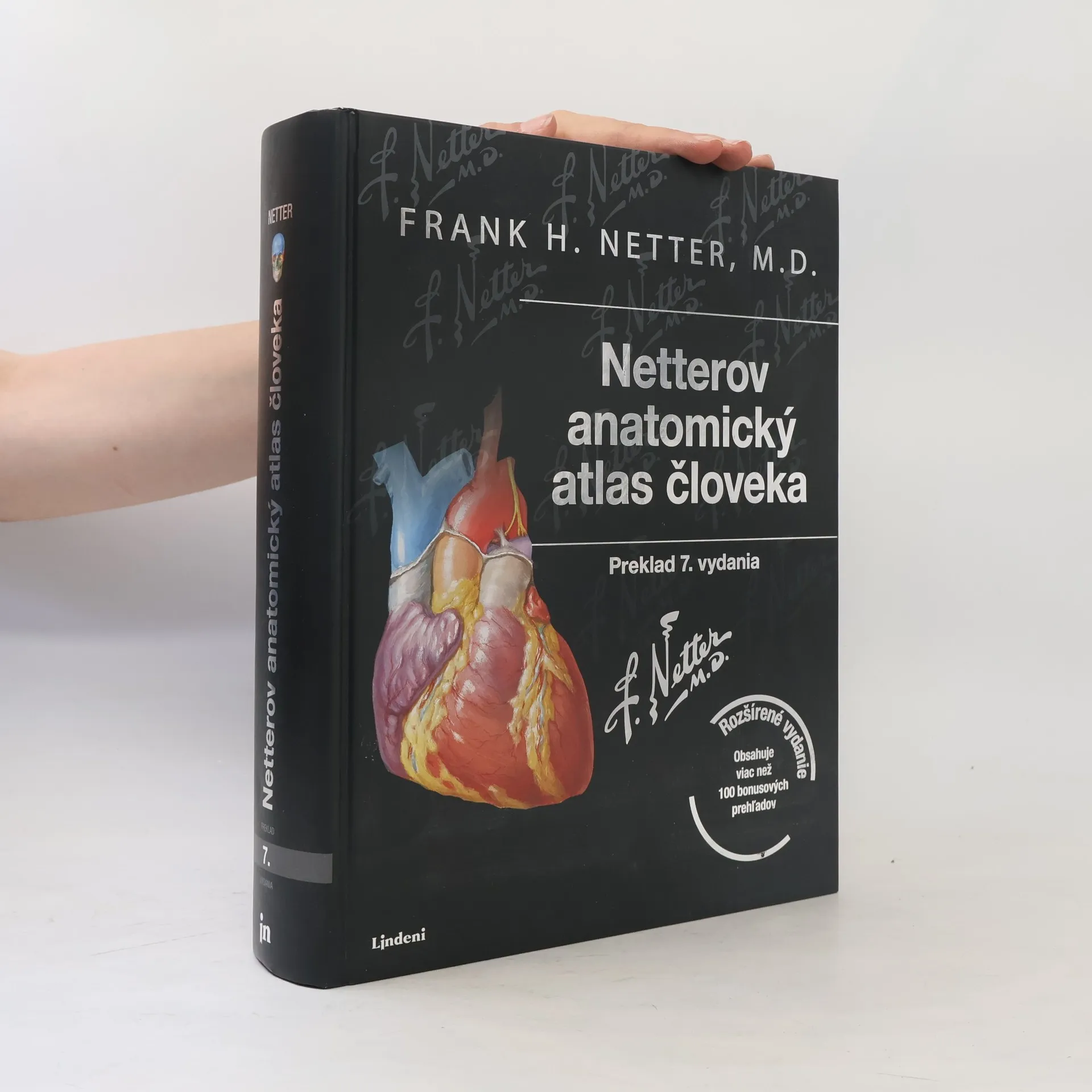
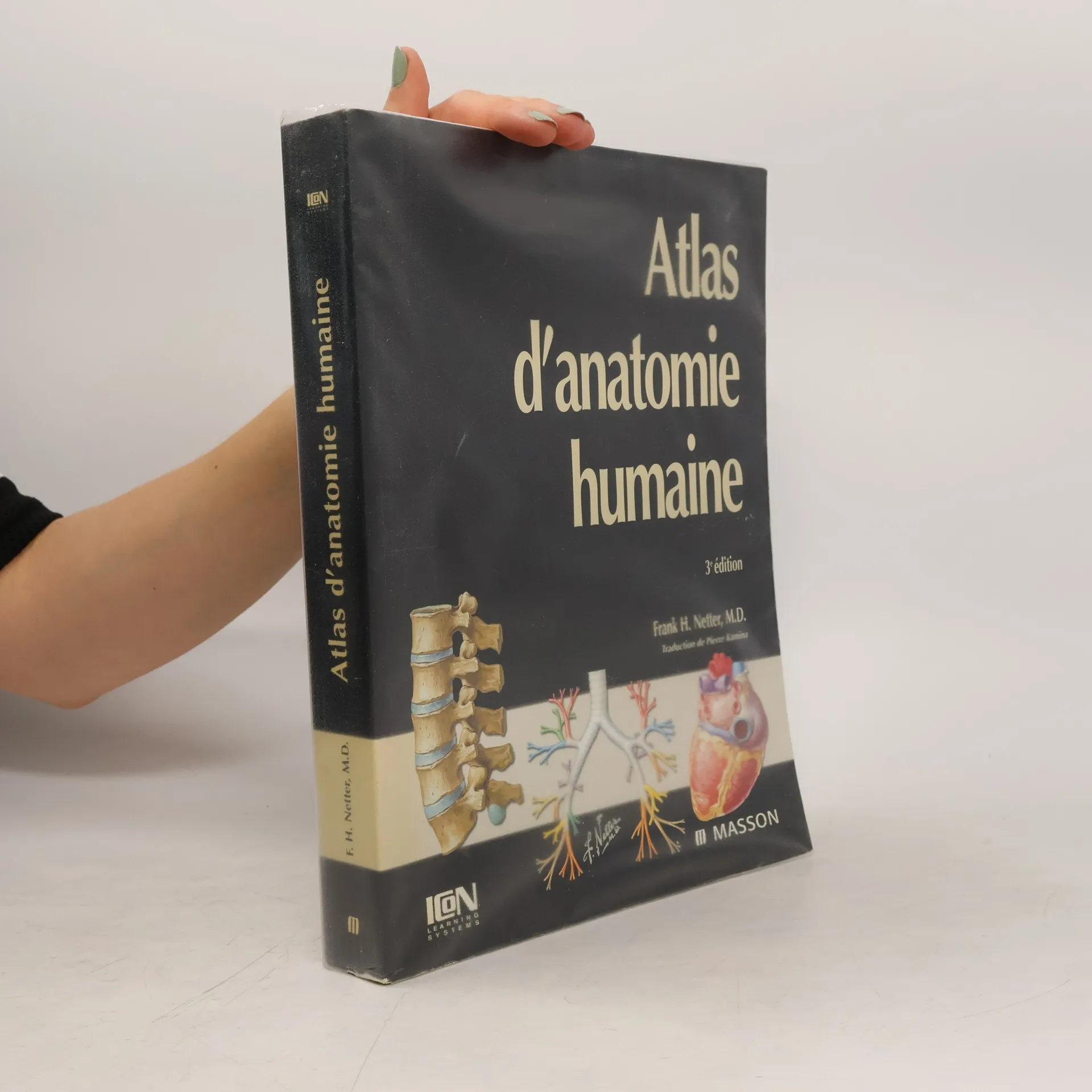
L'Atlas d'anatomie de Netter est un livre de référence internationale en raison de ses qualités iconographiques, scientifiques et pédagogiques. Les 900 magnifiques illustrations, qui rendent cet ouvrage si attrayant ne le cèdent en rien à l'exactitude scientifique. La sélection des figures et le choix des couleurs permettent au lecteur de saisir immédiatement l'élément qu'il recherche. Cette 3e édition s'est enrichie de 50 clichés d'imagerie moderne (TDM et IRM), et de schémas d'anatomie de surface, indispensables pour pouvoir aborder aisément la sémiologie et la pathologie. Les légendes, conformes à la terminologie internationale (Nomina Anatomica francisée), facilitent la compréhension et la mémorisation des termes anatomiques. Des commentaires didactiques viennent parfois éclairer le lecteur sur des points particuliers. L'ouvrage est divisé en huit sections, qui font chacune le tour complet d'une grande région anatomique. Ce livre rassemble donc clairement l'ensemble des connaissances anatomiques nécessaires à tous les étudiants, futurs médecins, dentistes, sages-femmes, kinésithérapeutes, infirmiers... Le " Netter " mérite vraiment une place de choix dans toute bibliothèque médicale.
Frank H. Netter Livres
Frank H. Netter était un médecin et un illustrateur médical de premier plan qui a uni les mondes de l'art et de la médecine. Ses dessins anatomiques détaillés et précis ont profondément influencé l'éducation et la pratique médicales. Il a utilisé son talent artistique pour créer des œuvres visuellement captivantes qui ont aidé d'innombrables étudiants et praticiens à comprendre les complexités du corps humain. Son héritage durable constitue une pierre angulaire de l'illustration médicale et de la clarté dans la compréhension anatomique.


Atlas of Human Anatomy 7
- 672pages
 - 24 heures de lecture
 
"The only anatomy atlas illustrated by physicians, Atlas of Human Anatomy, 7th edition, brings you world-renowned, exquisitely clear views of the human body with a clinical perspective. In addition to the famous work of Dr. Frank Netter, you'll also find nearly 100 paintings by Dr. Carlos A. G. Machado, one of today's foremost medical illustrators. Together, these two uniquely talented physician-artists highlight the most clinically relevant views of the human body. In addition, more than 50 carefully selected radiologic images help bridge illustrated anatomy to living anatomy as seen in everyday practice"--Publisher's description
The Ciba Collection of Medical Illustrations. Volume 4. Endocrine System and Selected Metabolic Diseases
- 303pages
 - 11 heures de lecture
 
Netter Atlas of Human Anatomy: Classic Regional Approach
paperback + eBook
- 556pages
 - 20 heures de lecture
 
Focusing on the needs of students and clinical professionals, this anatomy atlas offers clear, detailed illustrations of the human body, organized by region. Its unique approach highlights key anatomic relationships relevant to clinical practice, featuring over 550 meticulously crafted plates alongside selected radiologic images for common perspectives. Created by clinicians for clinicians, it serves as an essential resource for those learning anatomy, participating in dissection labs, or refreshing their knowledge.
With hundreds of illustrations, this title features informative text by medical experts; anatomy, physiology, and pathology; and diagnostic and surgical procedures.
Netter Atlas of Human Anatomy: Classic Regional Approach (hardcover)
Professional Edition with NetterReference Downloadable Image Bank
- 712pages
 - 25 heures de lecture
 
This atlas serves as a comprehensive resource for clinical professionals and students, offering detailed illustrations of human anatomy from a clinician's perspective. It features over 550 plates and radiologic images, highlighting key anatomical relationships essential for training and practice. New illustrations and nerve tables enhance coverage of clinically significant areas. The content is guided by expert anatomists and adheres to international anatomical standards, making it a vital tool for dissection labs and patient education. Additionally, it provides access to a downloadable image bank for personal use.
The Netter Atlas of Human Anatomy is an essential resource for students and clinical professionals. It offers detailed illustrations of the human body, emphasizing important anatomical relationships for clinicians. With over 550 exquisite plates and selected radiologic images, it serves as a comprehensive guide for learning and refreshing anatomy knowledge.
Netter's Head and Neck Anatomy for Dentistry
- 659pages
 - 24 heures de lecture
 
Netter's Head and Neck Anatomy for Dentistry, by Neil S. Norton, PhD, uses more than 600 full-color images from the Netter Collection to richly depict all of the key anatomy that's relevant to clinical practice. This new edition takes your knowledge further than ever with more Netter illustrations; addition of over 20 cone beam CT images; new chapters on the upper limbs, thorax, and abdomen; and more than 100 multiple-choice questions. Whether for your dental anatomy course, board review, or as a handy reference in your dental office, this concise, visual guide is an excellent anatomy atlas and quick reference for students and professionals in dentistry and dental hygiene. Identify clinically relevant anatomy with Netter illustrations highlighted and modified for dentistry. See the practical important of anatomy from illustrated clinical examples in each chapter. Review essential concepts easily with tables that display the maximum amount of information in an at-a-glance format. Master anatomy for the head and neck and beyond, including upper limbs, thorax, and abdomen. Stay current on hot topics like cone beam CT imaging, intraoral injections, and anesthesia. Recognize the context and clinical relevance of head and neck anatomy through additional coverage of dental procedures. Prepare effectively for the dental boards with over 100 multiple-choice questions.
Netter's Atlas of Human Embryology
- 267pages
 - 10 heures de lecture
 
Here's a rich pictorial review of normal and abnormal human prenatal development. For each body system or region, you'll find a brief description of the developmental plan, with key concepts and terminology, followed by discussions of histological principles, the classification of congenital defects, and basic cellular, molecular, and genetic concepts.An emphasis on morphological patterns in the embryo and fetus makes it easy to understand the structure and function of the adult body and the embryonic basis of birth defects.Summary tables and terminology sections at the end of each chapter, plus an appendix with all major congenital defects and their embryonic basis, make it easy to review course material and prepare for the USMLE.
The most critically acclaimed of all of Dr. Frank H. Netter's works, this 8-volume/13-book reference collection includes: hundreds of world-renowned illustrations by Frank H. Netter, MD; informative text by recognized medical experts; anatomy, physiology, and pathology; and diagnostic and surgical procedures.

