


Évaluation du livre
Paramètres
- 144pages
- 6 heures de lecture
En savoir plus sur le livre
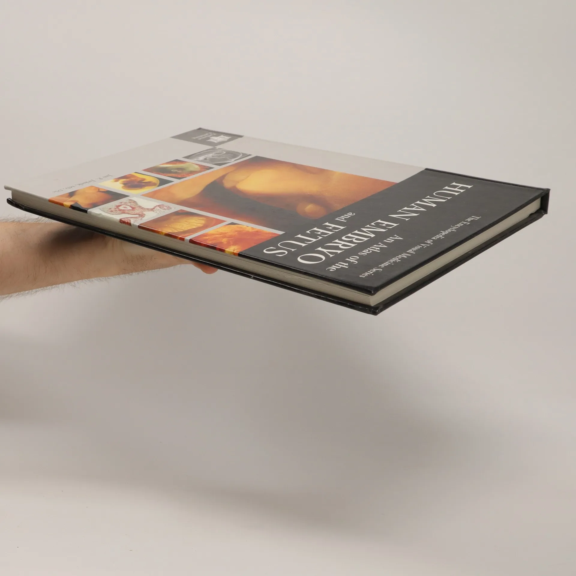
With hundreds of original photographs, optical micrographs and scanning electron micrographs, this atlas describes the progress of the embryo throughout its development, highlighting the formation and differentiation of organ structures. From the preembryonic and embryo stages to the development of the skeleton and striated muscle, organogenesis of the heart, and development of external genitalia, it provides authoritative answers to the most frequently asked question about the human embryo. With its plethora of outstanding photographs and images, experienced embryologists as well as clinicians and students can compare historical ideas with photographic reality.
Achat du livre
An Atlas of the Human Embryo and Fetus: A Photographic Review of Human Prenatal Development, Louis G. Keith, Jan Evangelista Jirásek
- Langue

- Année de publication
- 2000
- product-detail.submit-box.info.binding
- (rigide)
Modes de paiement
Il manque plus que ton avis ici.
- Titre
- An Atlas of the Human Embryo and Fetus: A Photographic Review of Human Prenatal Development
- Langue
- Anglais
- Éditeur
- Parthenon Publishing Group
- Publié
- 2000
- Format
- rigide
- Pages
- 144
- ISBN10
- 185070659x
- ISBN13
- 9781850706595
- Séries
- Évaluation
- 3,5 sur 5
- Description
- With hundreds of original photographs, optical micrographs and scanning electron micrographs, this atlas describes the progress of the embryo throughout its development, highlighting the formation and differentiation of organ structures. From the preembryonic and embryo stages to the development of the skeleton and striated muscle, organogenesis of the heart, and development of external genitalia, it provides authoritative answers to the most frequently asked question about the human embryo. With its plethora of outstanding photographs and images, experienced embryologists as well as clinicians and students can compare historical ideas with photographic reality.