
En savoir plus sur le livre
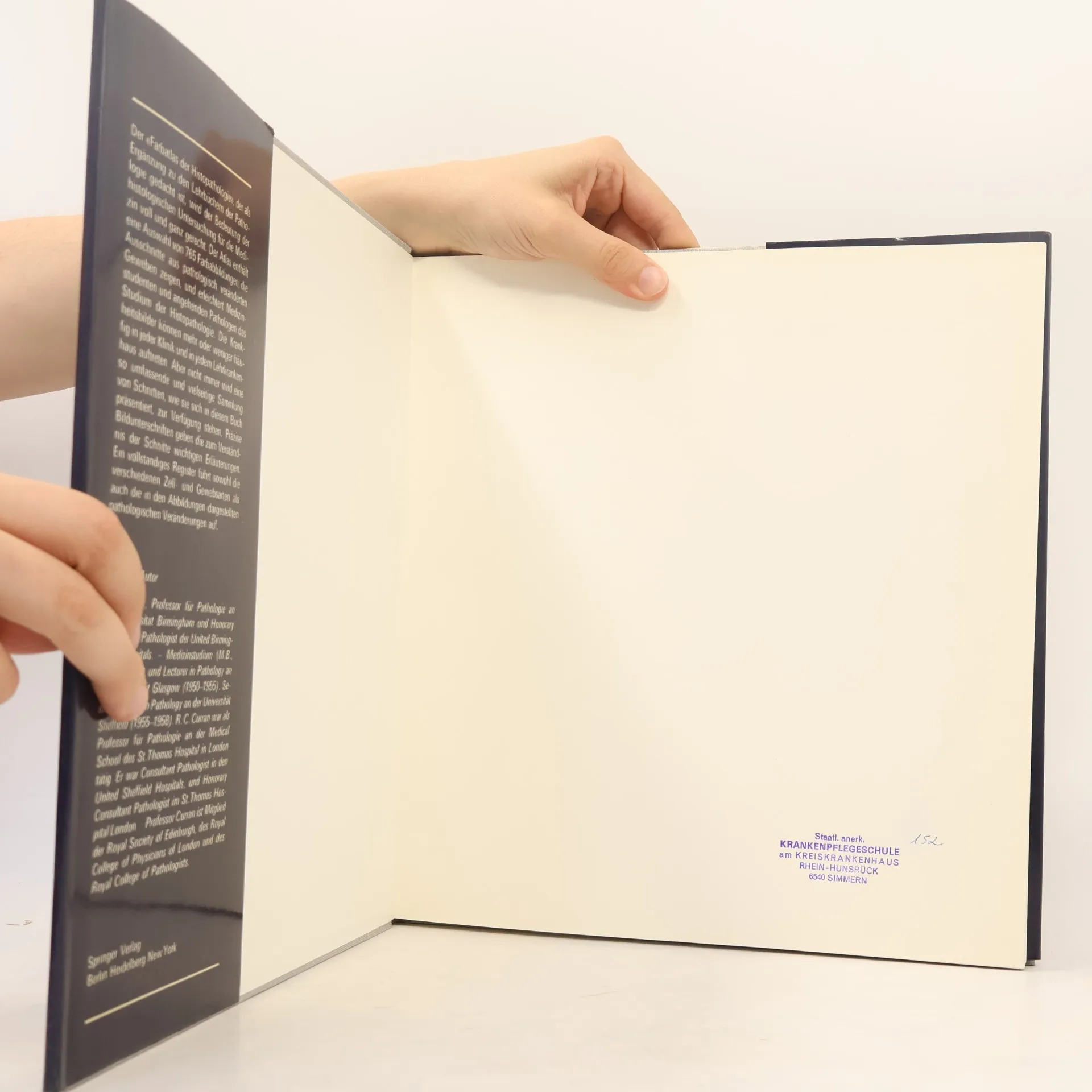
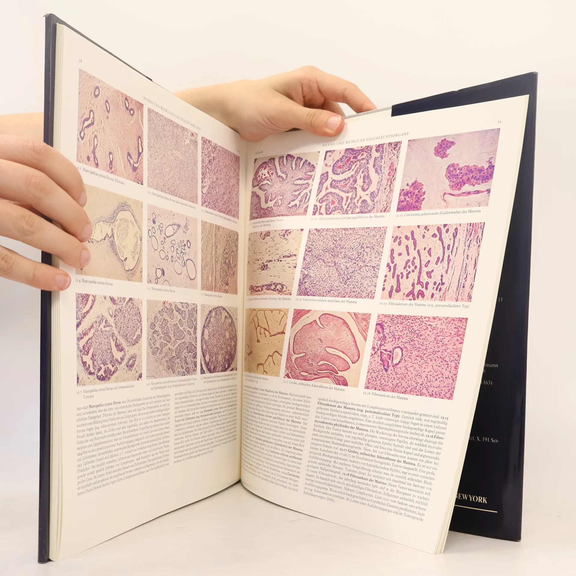
This fourth edition of Professor Curran's renowned colour atlas of histopathology features a fully revised text and additional immunohistological images, enhancing its 804 full-colour illustrations. The clarity of the text and the balanced colour of the sharp illustrations make this atlas an invaluable reference for both students and pathologists. Each illustration is meticulously selected for its structural significance, with precisely defined boundaries to minimize wasted space. The overall organization remains consistent, with chapters dedicated to the main systems or organs of the body. An introductory chapter covers essential tissue reactions in disease, equipping students with the foundational language of histopathology to interpret microscopic changes effectively. While most conditions depicted are common or fairly common diseases, some rare lesions are also included. This atlas primarily serves to convey visual information, complementing existing textbooks. A new comprehensive index enhances usability. Aimed mainly at undergraduate students, this edition is also likely to benefit postgraduate students training in pathology or other clinical fields, based on the success of its predecessors.
Achat du livre
Farbatlas der Histopathologie, Robert C Curran, K. Bürki, H. George Burkitt
- Langue

- Année de publication
- 1975
- product-detail.submit-box.info.binding
- (rigide),
- État du livre
- Abîmé
- Prix
- 18,56 €
Modes de paiement
Personne n'a encore évalué .
- Titre
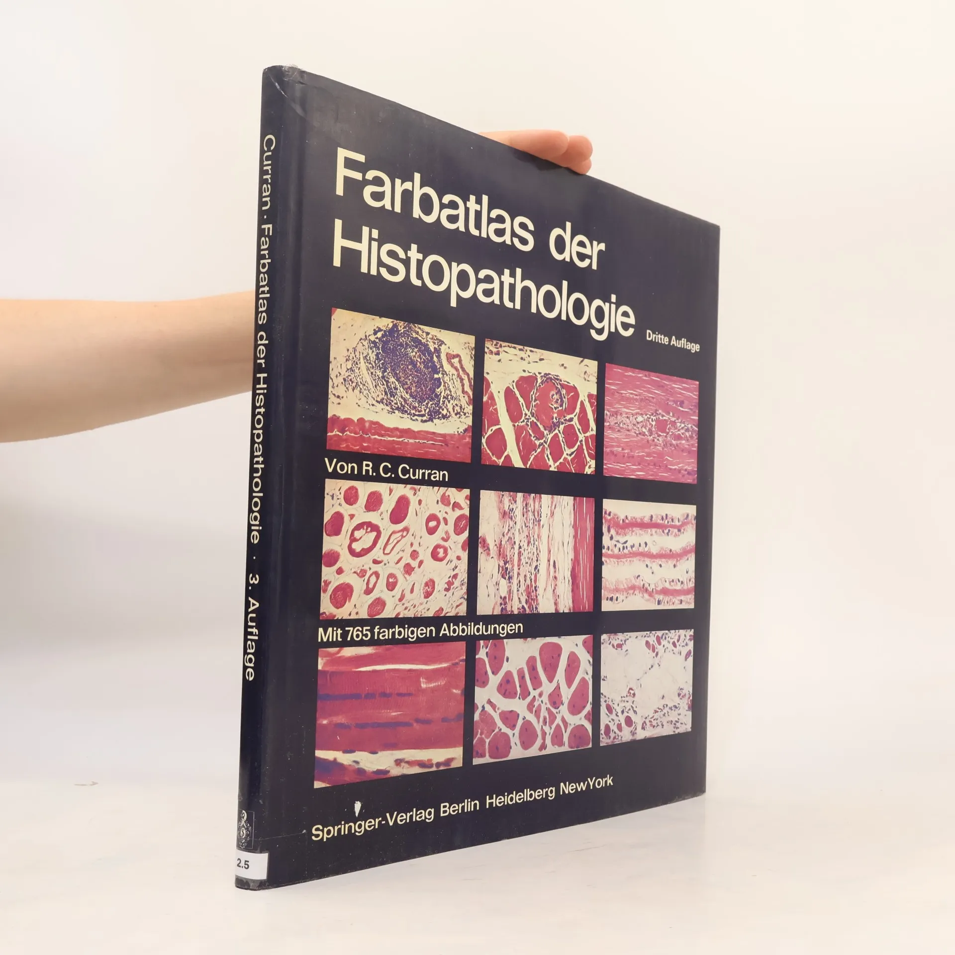
- Farbatlas der Histopathologie
- Sous-titre
- Mit 765 farbigen Abbildungen - Dritte Auflage
- Langue
- Allemand
- Auteurs
- Robert C Curran, K. Bürki, H. George Burkitt
- Éditeur
- Springer
- Publié
- 1975
- Format
- rigide
- Pages
- 96
- ISBN10
- 3540071911
- ISBN13
- 9783540071914
- Séries
- Mots clés
- Médecine, Pathologie
- Description
- This fourth edition of Professor Curran's renowned colour atlas of histopathology features a fully revised text and additional immunohistological images, enhancing its 804 full-colour illustrations. The clarity of the text and the balanced colour of the sharp illustrations make this atlas an invaluable reference for both students and pathologists. Each illustration is meticulously selected for its structural significance, with precisely defined boundaries to minimize wasted space. The overall organization remains consistent, with chapters dedicated to the main systems or organs of the body. An introductory chapter covers essential tissue reactions in disease, equipping students with the foundational language of histopathology to interpret microscopic changes effectively. While most conditions depicted are common or fairly common diseases, some rare lesions are also included. This atlas primarily serves to convey visual information, complementing existing textbooks. A new comprehensive index enhances usability. Aimed mainly at undergraduate students, this edition is also likely to benefit postgraduate students training in pathology or other clinical fields, based on the success of its predecessors.